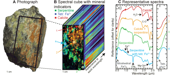
Figure 2.
Hyperspectral image of a serpentinite sample with red and green coatings (Nor4-14, described in Greenberger et al., 2015b) from Norbestos, Quebec, Canada. (A) Photograph of the full rock. (B) Image showing spectral parameters that map calcite (red), serpentine (green), and a feature at 0.45 µm (BD450; blue) due to tetrahedral Fe3+ within serpentine. The third dimension shows the reflectance as a function of wavelength for each pixel within the image, with black and purple being low and red high. (C) Plot with representative spectra of different units within the hyperspectral image. Colors correspond to colors in the spectral parameter image with locations numbered. Close-up views of the 0.45 µm feature are shown on the right. These images were acquired with Headwall Photonics Inc. High Efficiency Hyperspec® visible–near-infrared E-series (0.4–1.0 µm, 7 nm spectral resolution, 0.382 mrad instantaneous field of view) and High Efficiency Hyperspec® shortwave infrared X-series pushbroom systems (1.0–2.5 µm, 12 nm spectral resolution, 1.2 mrad instantaneous field of view) imaging spectrometers (see GSA Supplemental Data Repository [see footnote 1] for more information).