
Page 5 - gt1512
P. 5
Field- and lab-based Regional and global EXAMPLE 1: CHARACTERIZING HABITABILITY AND
imaging spectroscopy geologic units identified SEARCHING FOR BIOSIGNATURES IN SERPENTINE-
BEARING ROCK
Fine-scale units within
rocks identified The serpentinization process liberates molecular hydrogen that
can sustain microbial communities and react through biotic and
Headwall CAO- CRISM Mineral associations Basic compositional abiotic processes to form methane (e.g., Kelley et al., 2001, 2005;
AToMS Sleep et al., 2004; Oze and Sharma, 2005; Schulte et al., 2006;
hyperspectral OMEGA determined Cardace and Hoehler, 2009; Etiope and Sherwood Lollar, 2013;
M3 McCollom and Seewald, 2013). The oxidation states and coordi-
UCIS AVIRIS Hyperion nation environments of iron produce diagnostic absorption
features readily detected and mapped with imaging spectroscopy
Spectral bands HyMap but not so easily spatially resolved with traditional analysis tech-
niques. In serpentinized bodies, the oxidation state and iron coor-
multispectral WorldView-3 differences distinguished dination chemistry are related to the volume of hydrogen
produced and the availability of reduced gasses capable of
Mastcam/ ASTER supporting microbial metabolisms (Marcaillou et al., 2011;
Andreani et al., 2013). Though degrees of serpentinization are not
Pancam ThemLaisTnhVdesISmatis IRMODIS readily apparent visually, work by Greenberger et al. (2015b) used
HiRISE imaging spectroscopy in the 0.42–1.1 µm region to map the depth
of an electronic transition of tetrahedrally coordinated Fe3+ occur-
µm m km ring at 0.45 µm as a proxy for hydrogen production. Using new
imaging data that cover an extended wavelength range (0.40–2.5
Spatial resolution µm; Fig. 2), the 0.45 µm feature is shown with the two dominant
minerals, carbonate and serpentine, which exhibit sharp vibra-
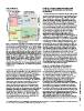
Figure 1. Conceptual plot of the relative spectral sampling versus the spatial tional absorptions mapped through calculation of the depths of
scales of observations by various imaging systems and the gap that ground- these features (Clark and Roush, 1984) and other spectral param-
based imaging spectroscopy fills (dashed box). Blue text indicates imaging eters (data processing algorithms are described in the GSA
systems on Earth, red italicized text is for Mars, and gray bolded text is for the Supplemental Data Repository1). Different portions of this sample
Moon. The number of spectral bands and the spatial resolutions used in this have undergone different degrees of serpentinization; those areas
plot are generally those in the visible-shortwave infrared regions. The Headwall with tetrahedral Fe3+ have undergone advanced serpentinization
and Ultra Compact Imaging Spectrometer (UCIS) imagers were used to and are promising areas to search with still higher spatial resolu-
acquire data presented in this paper. ASTER—Advanced Spaceborne Thermal tion compositional or isotopic techniques (e.g., scanning electron
Emission and Reflection Radiometer; AVIRIS—Airborne Visible/Infrared microscopy, mass spectrometry) for microbial biosignatures and
Imaging Spectrometer; CAO-AToMS—Carnegie Airborne Observatory– to understand the production of reduced gases.
Airborne Taxonomic Mapping System; CRISM—Compact Reconnaissance
Imaging Spectrometer for Mars; M3—Moon Mineralogy Mapper; OMEGA— EXAMPLE 2: HYDROTHERMAL ALTERATION AND
Observatoire pour la Minéralogie, l’Eau, les Glaces et l’Activité; HiRISE—High DIAGENESIS OF LACUSTRINE PILLOW BASALTS
Resolution Imaging Spectrometer Experiment; MODIS—Moderate Resolution
Imaging Spectrometer; THEMIS VIS/IR—Thermal Emission Imaging System Alteration rinds illuminate conditions of water-rock interac-
(visible/infrared). tions, and progressive changes from interior to exterior reflect
increasing degrees of alteration (e.g., Hausrath et al., 2008). With
mission (Van Gorp et al., 2014), and the MicrOmega instrument sub-millimeter spatial resolutions, imaging spectroscopy GSA TODAY | www.geosociety.org/gsatoday
will be on the upcoming ExoMars rover and Hayabusa-2 mission measurements of alteration rinds resolve fine changes in miner-
(Pilorget and Bibring, 2013). An orbital VSWIR imaging spec- alogy with alteration. Data from an Early Jurassic lacustrine
trometer has also been selected to fly to Europa. The shortwave pillow lava from the Hartford Basin are shown in Figure 3
infrared wavelengths are critical for mineralogic analyses (Greenberger et al., 2015a). In this work, coordinated imaging
because unique overtones and combination tones of vibrations spectroscopy, electron microprobe, microscopic X-ray diffraction,
within mineral structures occur in this region. The visible wave- microscopic thermal emission spectroscopy, and microscopic
lengths alone cannot distinguish mineralogies. While not the Raman spectroscopy analyses of a thick section across an altera-
focus of this paper, similar systems are in development to tion rind (Fig. 3C) characterized spectral, mineralogic, and chem-
measure thermal infrared emissivity in the laboratory (e.g., ical transformations. Unexpected key identifications with imaging
Edwards and Christensen, 2013). There is a steadily growing spectroscopy include calcic clinopyroxenes interpreted to have
suite of literature on the use of imaging spectroscopy of outcrops formed through >400 °C hydrothermal alteration (e.g., Bird et al.,
and samples, including core scanning, to answer geologically 1984; Manning and Bird, 1986), pervasive oxidation, Fe/
relevant questions (e.g., Kruse et al., 2012; Kurz et al., 2012; Mg-phyllosilicate signatures that increase in strength toward the
Murphy et al., 2012, 2014; Butz et al., 2015; Greenberger et al., exterior, and higher water content of calcites in the rind, either as
2015a, 2015b; Yokoyama et al., 2015). Here we highlight some
exciting applications of this emerging, non-destructive tech-
nology and the science discoveries enabled.
1 GSA supplemental data item 2015342, imaging spectroscopy methods and calibration, is online at www.geosociety.org/pubs/ft2015.htm. You can also request a copy
from GSA Today, P.O. Box 9140, Boulder, CO 80301-9140, USA; gsatoday@geosociety.org.
5